Union Biometrica provides flow cytometry for objects that are too large / too fragile for traditional cytometers and offer an alternative to manual sorting (under a microscope). These systems sort and dispense objects based on size and fluorescent parameters. Automating this process offers increased speed, sensitivity, quantification, and repeatability of experiments.
Systems for the automated analysis and sorting of viable multicellular organisms, cells and other large “particles” sized from 10–1,500 microns.
Learn More »
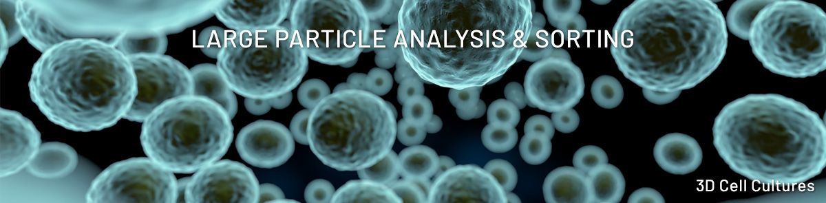
Union Biometrica, Inc.’s Large Particle Flow Cytometry technology is a high-throughput platform designed to analyze and sort intact 3D biological structures – hundreds of microns in size – without destroying them.
- Sort live organoids, spheroids, and small model organisms based on size, morphology, fluorescence, viability, or engineered reporters while keeping them intact and functional
Learn More »
- Scale from dozens to tens of thousands of organoids per run, accelerating screening, culturing processes, and QC workflows
- Standardize 3D biology with consistent, quantifiable inputs
- Eliminate subjective, manual picking, which leads to variability in organoid-based assays
Learn More »
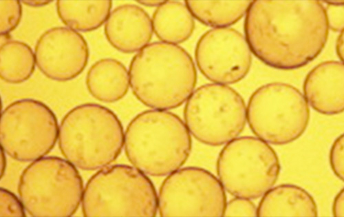
- Sort spheroids based on size (Time of Flight – TOF), optical density (Extinction), and Fluorescence
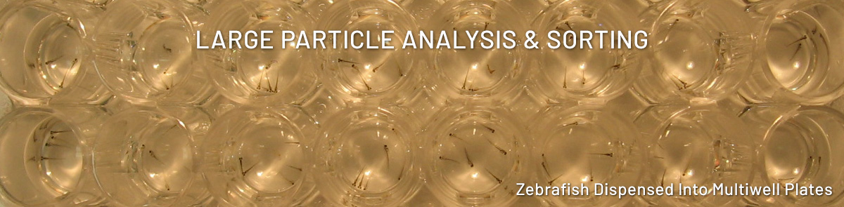
- Dispense GFP+ spheroids into multi-well plates
- Produce 96-well plates where every well contains one uniform GFP+ spheroid
Learn More »
Using the COPAS Vision™
- Acquire population-level and object-level data on organoid cultures
- Collect 1000s of brightfield images in flow
- Compare batches across days, conditions, and research sites
Learn More »
- The COPAS Vision™ captures brightfield images in flow
- Acquire cytometry, Profiler™, and image data at the same time
- Correlate phenotype to cytometry data
- Image analysis data include measurements of object size, width, height, smoothness and more!
Learn More »
- Blastocysts are structures that form early in embryogenesis shortly following fertilization
- Human blastocysts are made up of about 100-200 cells, they are typically 100-200 microns in diameter and can be analyzed on our COPAS instruments.
- The COPAS Vision equipped with a 500 micron flow cell was used to acquire cytometric data and brightfield image data from mouse blastocysts produced under various conditions as described in Dura et al. (EMBO J. 2025)
Learn More »
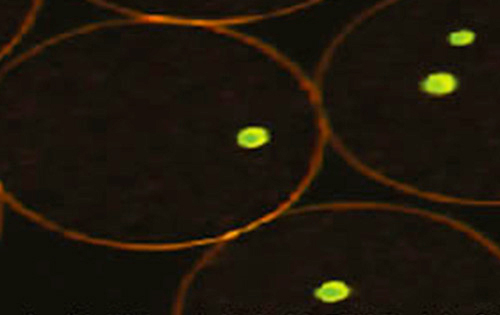
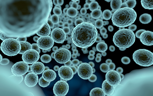
- Tumor cells grown in spheroids may provide more predictive models for tumor biology than monolayer cultures
- Profiler™ data generated by the COPAS Vision 1000 were used to characterize the localization of three cell types in cultured spheroids as described in Shah et al. (J. of Liposome Res. 2025)
- Brightfield images (inset) were captured in flow
Learn More »
- Picking induced pluripotent stem cell (iPSC) clusters remains largely manual and technique-dependent
- The COPAS FP-1000 was used to sort ~ 1mm iPSC clusters as described in Ma et al. (Front. Bioeng. Biotechnol. 2022)
- Pluripotency was preserved following cluster harvest, sorting, and passage
Learn More »
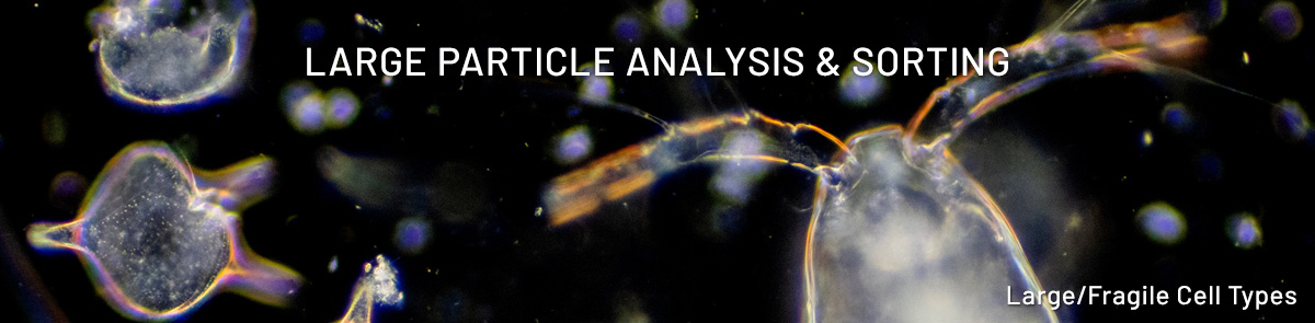
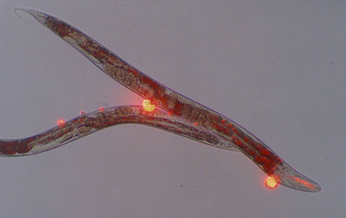
- Cardiomyocytes – and other cells that are long, skinny, and fragile – are challenging to sort using traditional FACS
- Large Particle Flow Cytometry is suited to carry out single cell sorting of viable and functional cardiomyocytes as described in Kannan et al. (Circulatory Research 2019)
- Enables single-cell transcriptomics of cells not amenable to traditional isolation methods
Learn More »
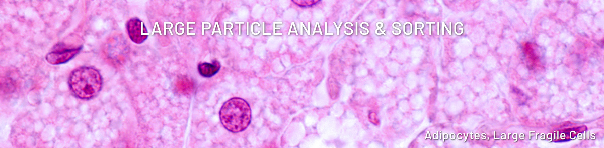
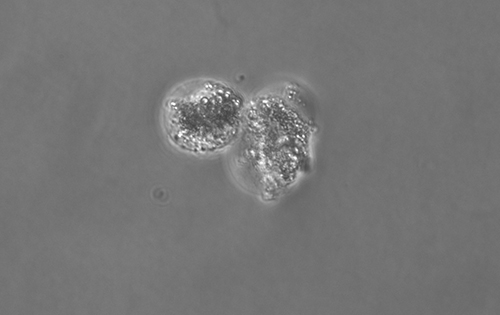
- Adipocytes – and cells that are large and prone to rupture – are challenging to sort using traditional FACS
- The BioSorter equipped with a 500 micron flow cell is suited to carry out single-cell sorting and analysis of adipocytes as described in Wang et al. (Nat. Commun. 2020)
- Enabled comparison of adipocyte size distribution between WT and KO mice
Learn More »
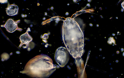
- Count, image, and identify organisms to measure biodiversity
- Marine plankton
- Freshwater meiofauna
- Soil micro- and meso-biota
- Sort organisms of interest for library creation, sequencing, or DNA barcoding
Learn More »
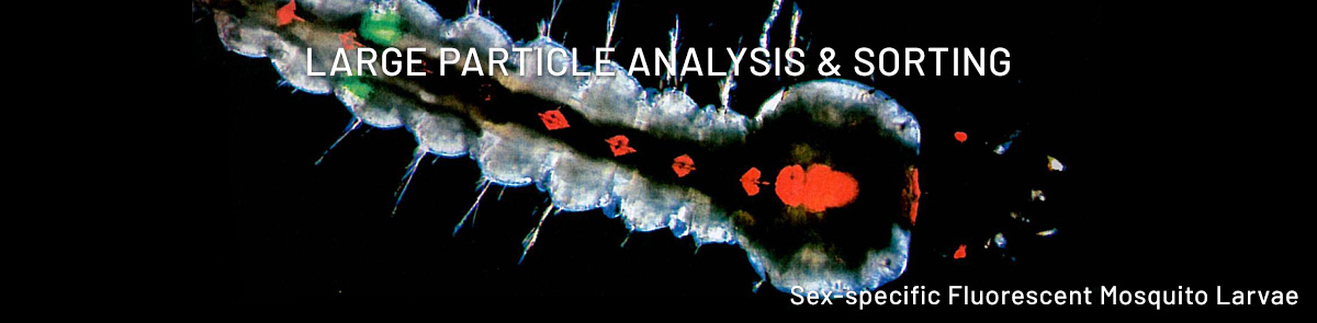
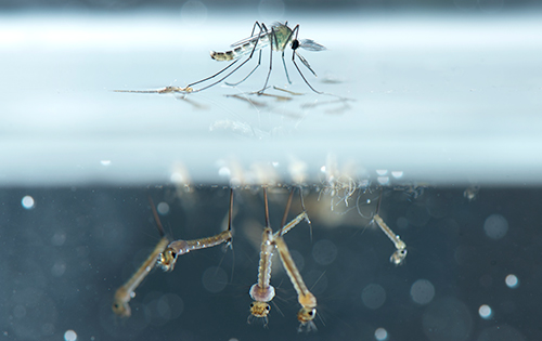
- Count and sort thousands of mosquito larvae
- Analyze and dispense populations based on fluorescence expression
- Enables accurate, large scale sex separation for Sterile Insect Technique (SIT) measures
Learn More »
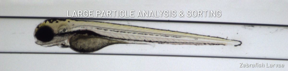
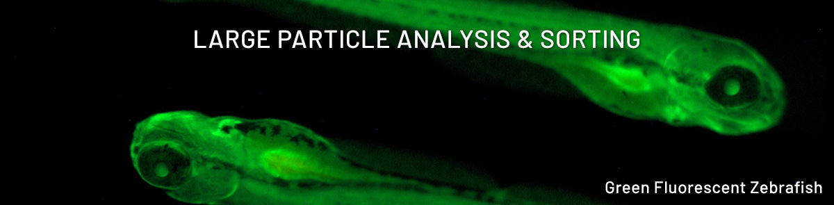
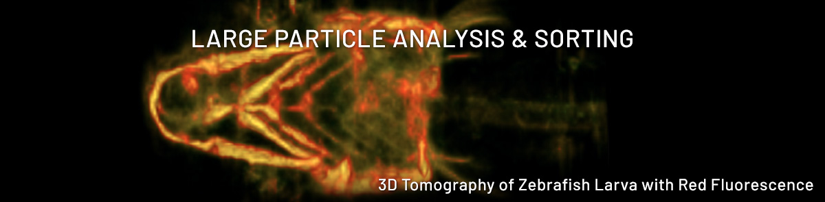
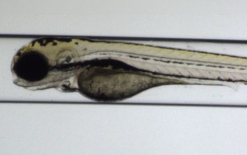
- The Vertebrate Automated Screening Technology (VAST) automates loading, orientation, and position 2-7 dpf zebrafish
- Image with on board camera for whole body/organ resolution
- Mount to a microscope for cellular resolution imaging
- Enables scale-up, improving reproducibility while reducing manual handling
Learn More »
- Large particle imaging flow cytometry enables quantitative analysis and precision dispensing of intact 3D models while preserving biological context.
- Integrated cytometric, imaging, and Profiler-based measurements support reproducible characterization of spheroid populations without dissociation.
- Cryopreserved spheroids can be distinguished from debris, analyzed at population scale, and dispensed into assay-ready multi-well plate formats.
- Drug response assays performed on intact cryopreserved spheroids demonstrate the utility of this platform for standardized and scalable 3D model workflows.
- These capabilities support the growing need for reproducible, high-throughput workflows aligned with NAM initiatives.
Learn More »
- Count and sort screwworm eggs and larvae for accurate quantification
- Analyze and dispense for assays that use multiwell plate formats
- Fluorescence measurements of transgenic variants with applications to SIT and pesticide screening
Learn More »