COPAS Flow Cytometers
COPAS Vision Cytometers
Imaging - Sorting - Cytometry!

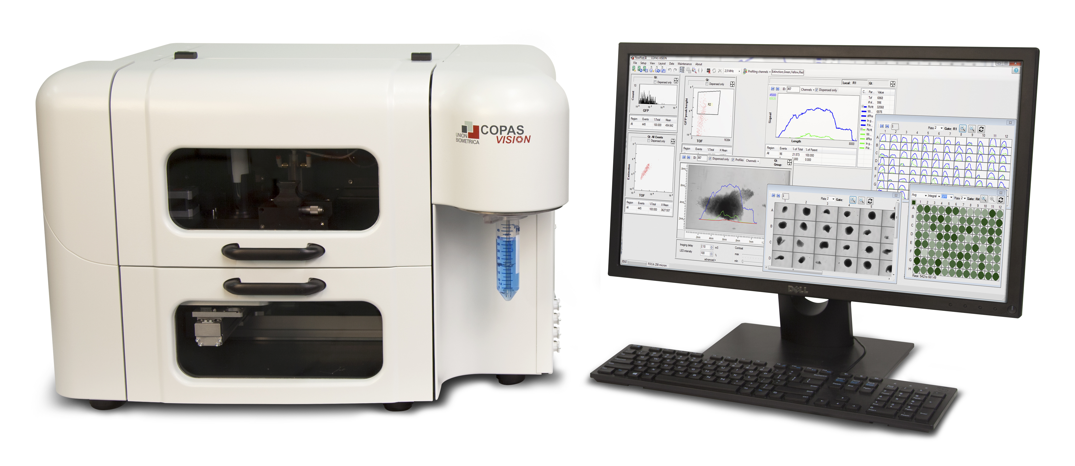
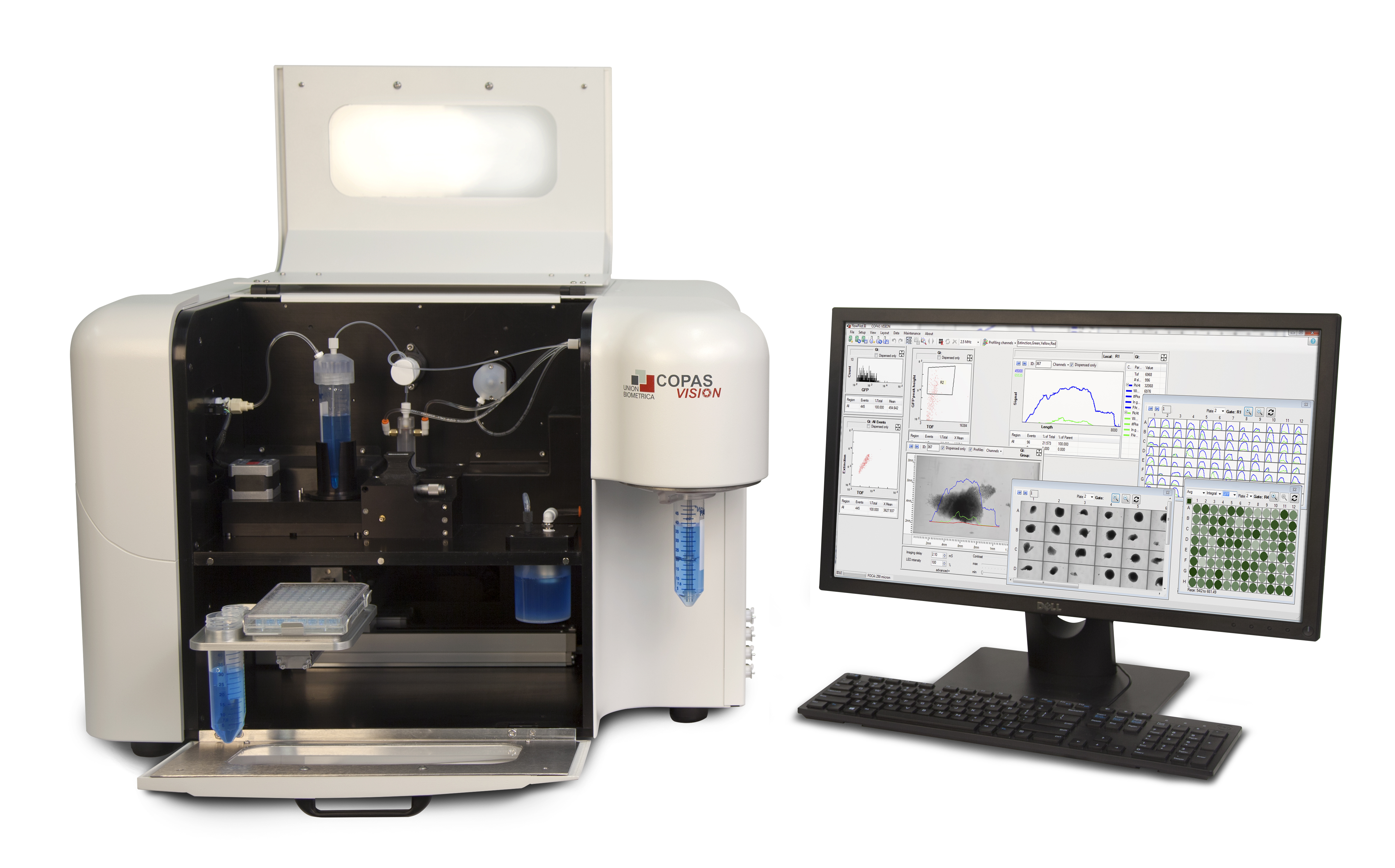
COPAS Vision adds real-time brightfield imaging to our large particle sorting capabilities. Instead of requiring a second step to move your samples to a separate microscope for post-run analysis, COPAS Vision allows you to quickly identify on the fly what objects/organisms are in a particular gate region, analyze count/concentration in a well, and observe sample morphology.
With COPAS Vision brightfield images of each object can be captured as it travels through the flow cell. These image data can be stored and correlated with Profiler data and other parameters. Up to 300 images per second can be captured with a resolution of 2.0 µm.
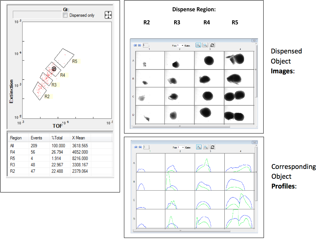
Cytometry and Imaging Data of Dispensed Organoids

Some examples of applications include:
- Large fragile cells (hepatocytes, cardiomyocytes, encapsulated cells)
- Cell clusters (organoids, spheroids, EBs, neurospheres, tumorspheres)
- Model organisms (C.elegans, zebrafish, marine samples)
- Plant/agricultural samples (Arabidopsis & tobacco seeds, parasitic nematodes, insect eggs & larvae)
Images overlaid with corresponding Profiler data:

Key Features:
- Choose from 4 models that span a range of sample diameters
COPAS Vision
flow cell:Object Size
RangeRecommended
object size250 µm 2 – 200 µm 10 – 175 µm 500 µm 6 – 400 µm 30 – 350 µm 1000 µm 15 – 850 µm 30 – 750 µm 2000 µm 40 – 1500 µm 100 – 1400 µm (Note these are general guidelines. Please talk to one of our Application Scientists about your specific project and sample requirements.)
- Options for up to 4 excitation lasers and 6 or 12 optical detectors/channels.
- Large objects are NOT homogeneous in terms of fluorescence or optical density. As each object passes through the flow cell the Profiler feature records/maps variations in these signal intensities as well as calculating values for peak height, peak width, peak count and relative position. Sorting and dispensing decisions are based on user-selectable values for these measurements.
- The patented air diverter gently sorts and dispenses fragile cells and live organisms without loss of viability.
- The X-Y stage accommodates collection in multiwell plates, tubes and various bulk receptacles.
- The OSIS, an oscillating sample introduction system for extremely delicate samples, can be used with disposable syringes for introducing fragile samples.
Vision Sorting Video


