VAST Imagers
VAST Platform Overview
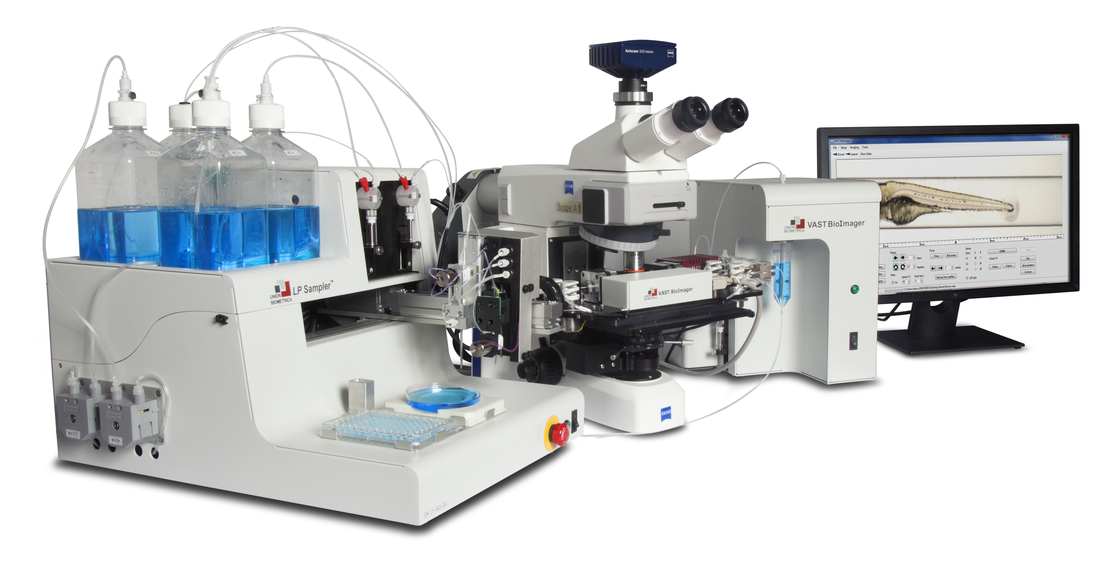
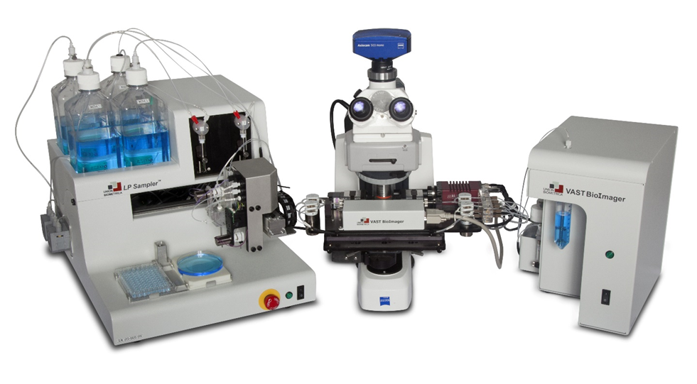
VAST BioImager – Whole Zebrafish Imaging and Positioning
The VAST BioImager system is for zebrafish researchers who are interested in high-resolution imaging of large numbers of 2–7 day old zebrafish larvae. The VAST BioImager automates the most demanding zebrafish handling, positioning, and orientation tasks. Using pattern recognition algorithms, a zebrafish larvae can be automatically loaded and positioned in both the desired lateral and angular orientations. This system can be mounted on upright microscopes to allow organ-level and cellular-resolution imaging tasks for high-throughput, high-content screens.
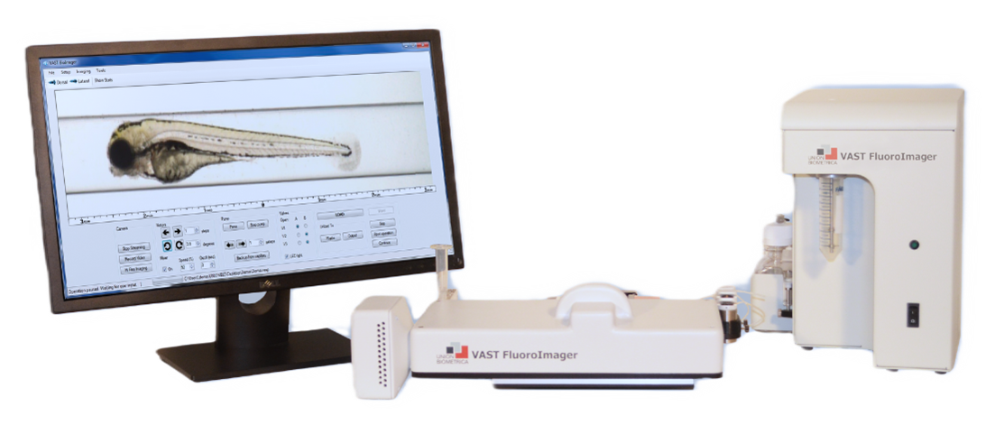
VAST FluoroImager – Automated Zebrafish Imaging with Fluorescence
The VAST FluoroImager system is for zebrafish researchers who are interested in brightfield and fluorescent imaging of large numbers of 2–7 day old zebrafish larvae. The VAST FluoroImager captures of 10-micron resolution, whole-fish, brightfield and fluorescent images in an automated and autonomous method. The VAST FluoroImager can generate multi-channel Optical Projection Tomography (OPT) datasets for 3D morphological analysis. Zebrafish larvae can be automatically loaded and positioned in both the desired lateral and angular orientations. This system can be mounted on a microscope to allow higher-magnification imaging tasks.
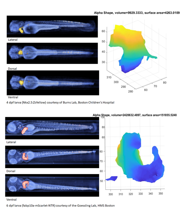
Optical Projection Tomography (OPT) with VASTomography
The VAST FluoroImager positions and rotates zebrafish larvae, allowing for an accurate reconstruction through the use of optical projection tomography (OPT). Along with the VASTomography software, the VAST can produce datasets used for quantitative 3D morphological analysis, advanced feature visualization, geometric calculation, and developmental studies.
Representative fully automatic WORK Flow process
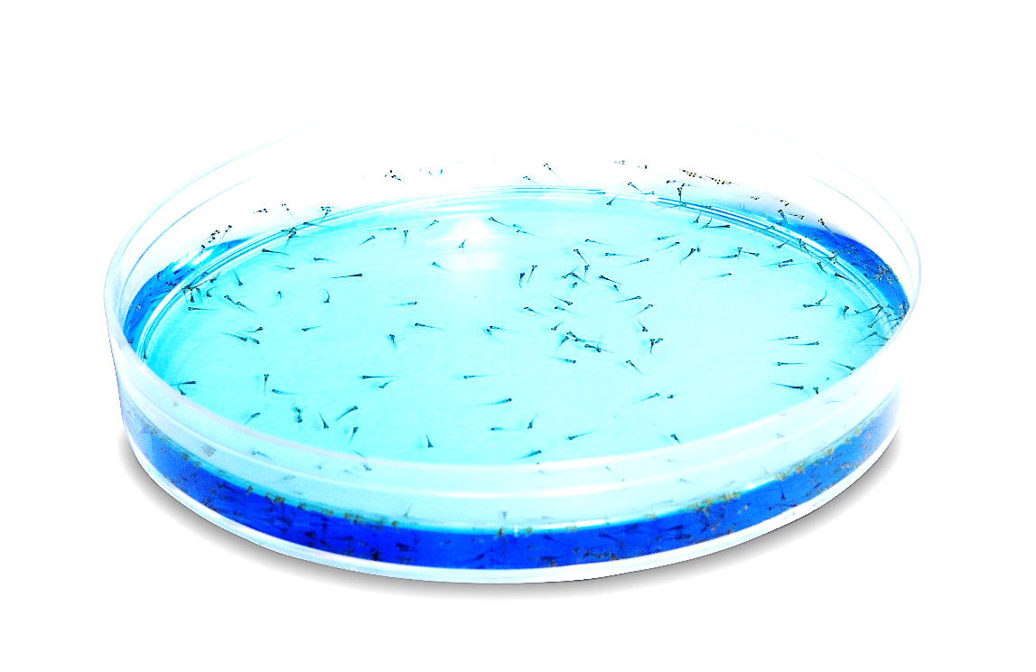
Larva is introduced from a sample source either from multiwell plates using the LP Sampler or from a bulk collection using the VAST Sample Cup or the VAST Pipettor. The VAST system automatically moves the larva into the imaging capillary.

The VAST performs an orientation check and initiates collection of images according to the activated jobs list from its onboard camera. Imaging settings can be configured to include single or multiple rotational angles, a focal depth series, video, and optical projection tomography and utilizing four different light sources to collect multiple brightfield and fluorescent images and videos.

After VAST imaging jobs are finished the system can initiate an External Imaging process. The VAST system will orient and position the larva to an optimal location for your high-content microscope's objective and then send a signal to the your microscope’s camera to take high-content pictures. The VAST system will then wait to receive a return signal which tells it to move to its next external imaging commands.

After all imaging is completed, VAST will output the fish. If using the LP Sampler & Dispenser, the fish will be dispensed to a position in a multiwell plate.

