VAST Imagers
Imaging & Tomography Software


VAST Software
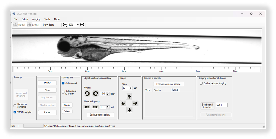
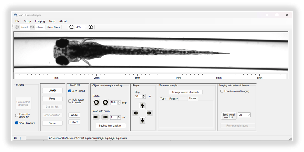
The VAST BioImager automates the process to take whole-fish brightfield images and to load, orient and position zebrafish larva at the focal point of an external microscope. The VAST FluoroImager expands upon this functionality incorporating the ability to collect brightfield and three colors of fluorescence images without the need for an external microscope.
VAST software follows a list of user-determined imaging collections
- Single or multiple rotational angle images (TIFF, JPEG, PNG)
- A depth series of images collected while the camera is focused on different depths of the capillary
- A short video (.avi)
- A compilation of images during rotational orientation that are used for 3D tomographic reconstruction
For each imaging collection, the user can customize the combination of excitation LEDs and emissions filters for optimal imaging of fluorescence expressed in the larva.
VASTomography™ software enables 3D Zebrafish Research
The VASTomography software can be utilized to compile the collection of images to make a 3D map of the lava to analyze volume and surface areas of expression.
- Ability to enerate 3D optical projection tomography (OPT) in minutes
- Data sets of volume, shape and position for selected region of interest
- 3D visualization with VolView and ParaView
- Data processing with MATLAB and Python
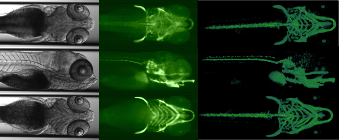
VASTomography software reconstruction and alpha Shape analysis of fluorescence characteristics.
The VASTomography software is a form of optical projection tomographic reconstruction based on light transmission through the sample. It transforms the rotational images collected by the VAST into a format that can be visualized and processed in 3D.
Characteristics such as volume and surface area of a region of interest can be calculated using the ‘alpha shape’ feature. All of which offers reliabe collection of images for positional, volumetric and shape information collected from the VAST FluoroImager or for higher content, from camera equipped to your upright higher resolution high microscope.
 Constructing tomography from fluorescent images of zebrafish larvae strain Sox10: Kaede. Images show brightfield, fluorescence and VASTomographic reconstruction for A) Dorsal view, b) Lateral view. C) Ventral view. Images captured by Zeiss AxioExaminer with a 5x lens / Axiocam 503 mono focused on VAST capillary.

