Our Technology
Zebrafish Imaging
Union Biometrica continues to develop new tools for model organism research. As a member of the zebrafish research community, we introduced this novel instrument platform for imaging zebrafish in early 2013. Based on technology from the Yanik Lab at MIT, the Vertebrate Automated Screening Technology (VAST), allows even the most demanding zebrafish handling and imaging tasks for automating high-throughput and high-content screens. This platform is designed for zebrafish researchers who are interested in imaging of large numbers of 2–7 day old zebrafish larvae.
Fully-automated orientation of zebrafish larvae

Reproduced from: Lab Chip, 2012, 12 711-716, Tsung-Yao Chang, Carlos Pardo-Martin, Amin Allalou, Carolina Wählby and Mehmet Fatih Yanik
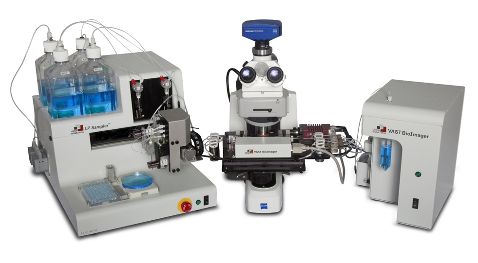
VAST BioImager – Whole Zebrafish Imaging and Positioning
The VAST BioImager system is for zebrafish researchers who are interested in high-resolution imaging of large numbers of 2-7 day old zebrafish larvae. The VAST BioImager automates the most demanding zebrafish handling, positioning, and orientation tasks. Using pattern recognition algorithms, a zebrafish larvae can be automatically loaded and positioned in both the desired lateral and angular orientations. This system can be mounted on upright microscopes to allow organ-level and cellular-resolution imaging tasks for high-throughput, high-content screens.

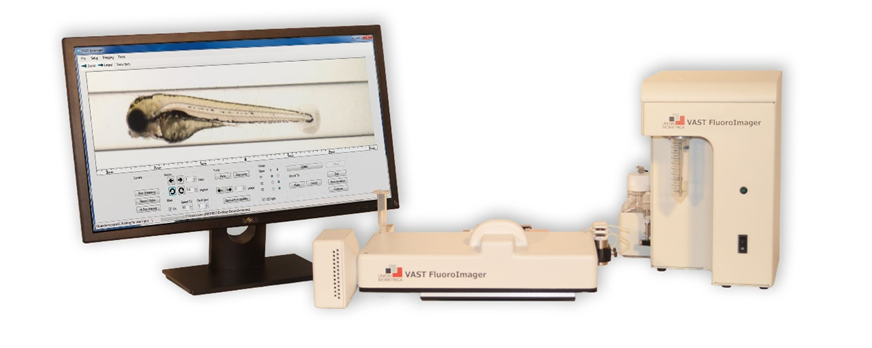
VAST FluoroImager – Automated Zebrafish Imaging with Fluorescence
The VAST FluoroImager system is for zebrafish researchers who are interested in brightfield and fluorescent imaging of large numbers of 2-7 day old zebrafish larvae. The VAST FluoroImager captures of 10-micron resolution, whole-fish, brightfield and fluorescent images in an automated and autonomous method. The VAST FluoroImager can generate multi-channel Optical Projection Tomography (OPT) datasets for 3D morphological analysis. Zebrafish larvae can be automatically loaded and positioned in both the desired lateral and angular orientations. This system can be mounted on a microscope to allow higher-magnification imaging tasks.

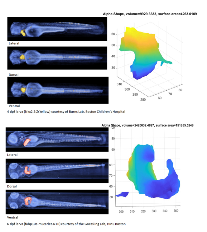
Optical Projection Tomography (OPT) with VASTomography
The VAST FluoroImager positions and rotates zebrafish larvae, allowing for an accurate reconstruction through the use of optical projection tomography (OPT). Along with the VASTomography software, the VAST can produce datasets used for quantitative 3D morphological analysis, advanced feature visualization, geometric calculation, and developmental studies. The Large Particle (LP) Sampler is an automated sample introduction system designed specifically for gentle handling of large fragile objects including large cells/clusters and model organisms such as zebrafish larvae. It is capable of aspirating samples from the wells of multiwell plates and delivering them intact to Union Biometrica’s COPAS, BioSorter and VAST systems. Software for seamlessly integrating the LP Sampler with these other systems is included. Sample object size can range from 1 to 1500 microns in diameter. This high-throughput device is capable of processing multiple samples simultaneously for continuous sample introduction. 
Autosampler Option

